Sunday, October 25, 2009

Serum Protein Electrophoresis

Tng Wess Lee, 0702570C
Clinical Biochemistry

Confirmation of Mband (Identifying Artefacts in SPE).

Serum Protein Electrophoresis (SPE) is a laboratory technique used to separate serum protein into 5 distinct fractions ( albumin, alpha 1, alpha 2, beta, gamma ). It can be used to measure the concentration of the serum protein as well. It can also be used to determine absence of normal proteins and to identify presence of abnormal proteins. SPE is normally requested for myeloma monitoring. It is often requested to identify M-band.

M-band, is a protein which is either immunoglobin or immunoglobin light chains. It will form a distinct band often at the gamma region. M-band is due to excessive production of immunoglobins due to diseases such as multiple myeloma, Monoclonal Gammopathy of Undetermined Significance, chronic lymphocytic leukemia, etc.
However, it is critical to ensure that the M-band seen on the SPE film is of an immunoglobin class. Sometimes, it may be due to artefacts. One good example of an artefact is fibrin frmo a plasma sample which looks like a M-band.

We therefore run immunofixation (IFE) to identify the immunoglobin class of the M-band.

Here are some pictures to illustrate what i have just said.
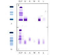


As you can see, the top blue bands are SPE of a sample which has a M-band. Confirmation from the IFE (purple) shows that the immunoglobin class is IgG/Kappa. However, the bottom blue bands of the SPE, is a plasma sample containing fibrin. It also has a distinct band in the gamma region which looks like a M-band. However when IFE is done, you notice that no distinct band of immunoglobin class appears. Therefore we understand that it is an artefact.

Saturday, October 24, 2009

Sherman: Molecular work

RNA extraction and quantification

The method of extracting and purifying RNA, which will be used for molecular studies

It involves harvesting cells, disrupting cells, homogenizing cells, bind RNA, collect RNA and wash membrane


Materials

  1. Rneasy-free pipette tips (filter tips)
  2. Microcentrifuge
  3. 96-100% and 70% non-denatured EtOH
  4. QIA shredder spin column
  5. Rnase spin column
  6. Pipette
  7. Micropipette
  8. Falcon tube
  9. Light microscope
  10. Counter
  11. RNA extraction kit
    1. RLT
    2. RW1
    3. RPE
  12. MQ water
  13. Cuvettes
  14. Spectrophotometer
  15. Centrifuge


Method

  1. Harvest cells (trypsinisng with treatment & transferring into a falcon tube for centrifuge)
  2. Centrifuge at 600rpm for 5mins, remove supernatant and count cells
  3. Centrifuge the remaining cells and remove the supernatant
  4. Disrupt the cells; on the cell pellets, add 350ul of RLT if the cell count is <>6 cells, or 600ul of RLT for 5 x 10⁶-107. Mix well (an alternative is to vortex for 1min)
  5. Homogenise cells; pipette lysate into QIA shredder spin column in 2ml collection tube (the shredder spin column will cut and lyse the cells more evenly to allow higher RNA yield)
  6. Centrifuge for 2mins at full speed and discard column (the RNA is in the flow-through)
  7. Bind RNA; add 1 volume of 70% EtOH to lysate (flow-through) and mix by pipetting - to precipitate the RNA

* 1 volume = amount of RLT that was added at the given cell count

  1. Collect RNA; transfer 700ul of sample into Rnase spin column in 2ml collection tube - RNA will be trapped in the membrane of the Rnase spin column
  2. Centrifuge for 15secs at full speed. Discard the flow-through
  3. Wash membrane; add 700ul of RW1 to the spin column, repeat the step above
  4. Add 500ul of RPE to the spin column, repeat the step above
  5. Repeat the step above, but centrifuge for 2mins (dry the RNA)
  6. Elute RNA; Transfer the spin column into an eppendorf tube
  7. Add 30ul of Rnase-free water (directly in the middle of the membrane, without touching it)
  8. Centrifuge at max speed for 1min
  9. Repeat the step above, recycling the Rnase-free water. This is to get higher yield of RNA
  10. Collect the flow-through.
  11. Label 5 baby eppendorf tubes for the storage of RNA (stored at -80ºc)
  12. Aliquote 5ul of RNA into the first 4 tubes, and 10ul into the last tube for RNA quantification

RNA quantitation

An absorbance of 1 unit at 260nm corresponds to 40ug of RNA per ml

Aλ260 = 1, concentration = 40ug/ml. To ensure significance, reading should be greater than 0.15

  1. Pipette 2ul of RNA into a cuvette and 498ul of MQ water

*Ensure that the RNA is mixed with the MQ water, otherwise there will be little yield reading

  1. Set up a blank sample (500ul MQ water) and the diluted RNA sample
  2. Insert the cuvettes into the spectrophotometer
  3. Set the wavelength (A260 and A280)
  4. Set reference for the blank to 0.000 absorbance
  5. [RUN] samples according
  6. Take readings for each cell line at the 2 given wavelengths (A260 and A280)

Purity of the RNA is measured by dividing the wavelength at A260 by A280. A ratio of 1.8 - 2.0 will give a pure result

Sunday, October 18, 2009

Rapid test for Dengue Virus,

Dengue virus is a mosquito-borne flavivirus and as everyone knows rapid detection and diagnosis is important. Rapid test kits are being introduced as it is able to detect and provide the result faster than routine laboratory methods like ELISA or Haemagglutination inhibition test.

Dengue Virus IgM / IgG antibody Rapid test kit.


Procedure.

  1. pipette 10 ul of patient serum into the square well
  2. drop 3 - 4 drops of test buffer into the round well
  3. start timer ( 15-20 mins )
  4. read results at 15 - 20mins
  5. results should not be read after 20 mins as false results might be obtained.

Reading of Results.

Purple or blue line should be observed at the C - control


If no line is observed, the test is invalid and have to be repeated.

Positive - purple or blue line observed at either M ( IgM ) or G ( IgG ) and C ( control )


Negative - only control line is observed.


Interpretation.


Positive IgM suggest primary or recent dengue infection.


Positive IgM &/or IgG suggest secondary or subsequent dengue infections.


Kenneth.
TG02